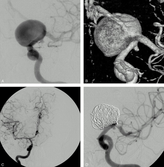
Fig 4.
SAH in a 64-year-old woman.
Conventional (A) and 3D (B) angiograms show a 20-mm aneurysm of the left ICA bifurcation with the A1 segment arising from the sac.
C, A balloon test occlusion shows the efficient collateral circulation through the AcomA.
D, Conventional angiogram at the end of the embolization shows a complete aneurysm occlusion and the sacrifice of the origin of the left A1 segment.

