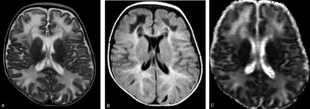
Fig 1.
Images obtained before dietary treatment. Axial T2-weighted image (A) showing hyperintense bilateral diffuse white matter involvement reaching the “U” fibers. The internal capsules are spared. The lesions are hyperintense with hypointense areas on FLAIR images (B). ADC map (C) shows increased diffusion in the white matter lesions.

