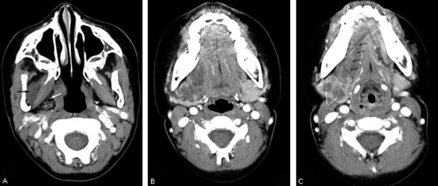
Fig 1.
Sequential images from axial contrast-enhanced CT scan demonstrating the plexiform neurofibroma of the right submandibular gland.
A–C, Tubular hypoattenuated masses are seen in the right masticator space (A, black arrow), parapharyngeal space (A, white arrow) and in the right submandibular space surrounding the submandibular gland. The right submandibular gland is enlarged and has peripheral enhancing parenchyma similar to that of the normal left submandibular gland (B, C). There is a central branching hypoattenuated mass (C, white arrows), which is the infiltrating plexiform neurofibroma. Note the extension of the plexiform neurofibroma into the right sublingual space along the course of the lingual nerve (C, black arrows).

