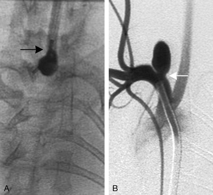
Fig 2.
Group 2 subject (high balloon position).
A, AP spot film obtained during aneurysm creation surgery. The RCCA is opacified with the elastase/iodinated contrast mixture. Note that a small portion of the inflated balloon has herniated into the proximal RCCA (black arrow).
B, Intra-arterial digital subtraction angiogram, right anterior oblique view, demonstrating a narrow-neck aneurysm (white arrow). Neck size is 1.9 mm.

