
Fig 2.
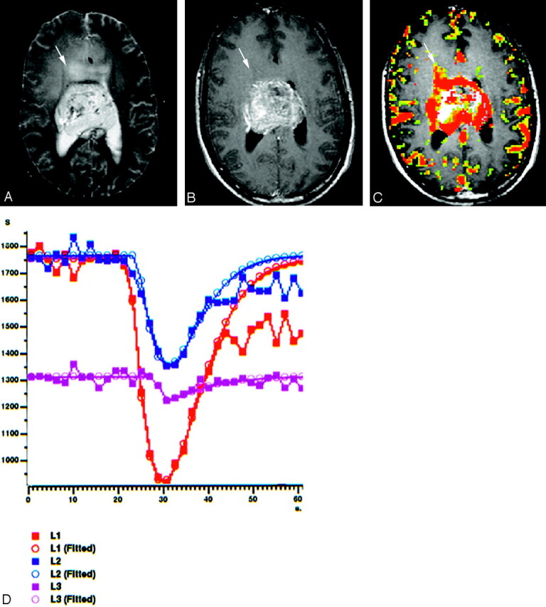
A 22-year-old man with centrally located glioblastoma multiforme (WHO IV).
A, Tumor shows heterogeneous hyperintensity with prominent peritumoral edema and/or tumoral infiltration (arrow) on axial T2-weighted SE image (2295/90).
B, There is a significant heterogeneous enhancement in tumoral borders but not in peritumoral area (arrow) on axial T1-weighted image (583/15).
C, Gradient-echo axial perfusion MR image (627/30) with rCBV color overlay map shows both high rCBVT value of 6.58 and rCBVP value of 2.21, which are consistent with HGGT. Peritumoral increased rCBV (arrow) shows tumoral infiltration outside the tumoral margins, which is not perceptible on T2- and contrast-enhanced T1-weighted images.
D, Time-signal intensity and gamma-variate fitted curves from tumoral (red), peritumoral (blue), and normal (purple) areas show prominent decrease in signal intensity from tumoral and peritumoral areas, when compared with signal intensity of normal gray matter.