
Fig 3.
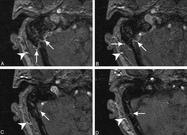
3D time-of-flight MR angiograms depicting the prominent right occipital artery (arrowhead) and branches permeating through the squamous portion of the occipital bone (arrows), bypassing the previously noted venous stenosis. Note abnormal nonsaturated flow in the dural venous sinus (arrowhead; images superior to inferior).