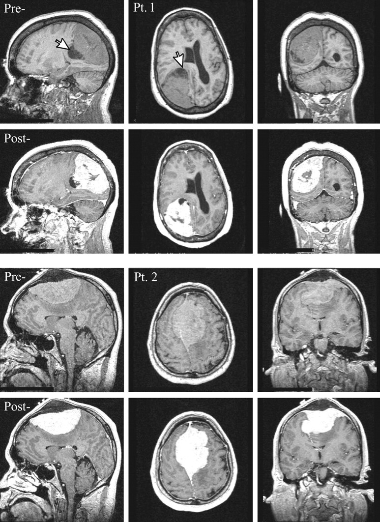
Fig 1.

Top, Sagittal, axial and coronal T1-weighted images pre- and postcontrast for patient 1. Note the large (6 × 5 × 5 cm3) extra-axial lesion in the right parietal region, surrounding small cysts (largest ∼1.5-cm diameter, arrow) and marked mass effect on the right lateral ventricle, midline shift, and sulcal effacement.
Bottom, Same for Patient 2. Note the large (9 × 6 × 4 cm3) lesion with its broad dural tail superiorly in the midline extending bilaterally through the falx and into the calvaria, homogenous enhancement, and mass effect reflected in displacement of both lateral ventricles and corpus callosum with moderate left-to-right midline shift.
