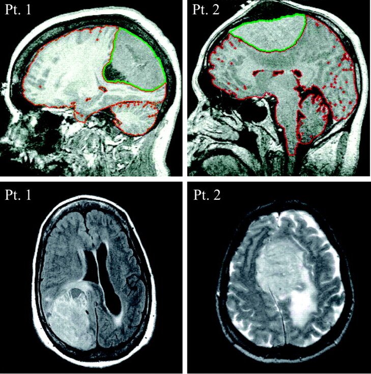
Fig 2.

Top, Sagittal T1 for patients 1 and 2. The red sulcal and ventricular outline is tissue/CSF segmentation. The extra-axial mass (outlined in green) is excluded from initial brain volume but then included to approximate preneoplastic VB.
Bottom, Axial fluid-attenuated inversion recovery and T2-weighted image for patients 1 and 2, respectively. In patient 2, note the considerable T2 hyperintensity, consistent with vasogenic edema, in the underlying parenchyma, particularly in the left centrum semiovale and parietal lobe.
