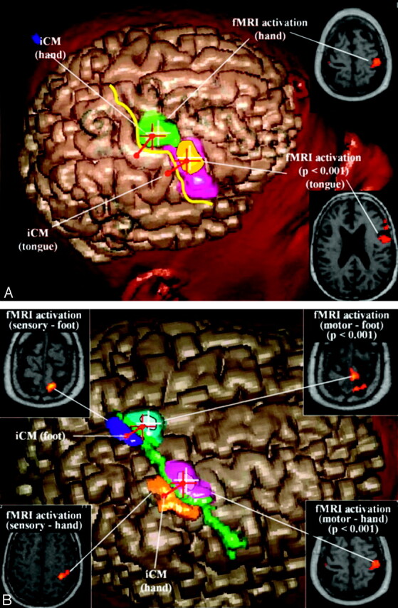
Fig 1.

A, Virtual 3D reconstruction (cortex surfacing method) of the right hemisphere in the navigation workstation showing the integration of data from iCM and fMRI in the case of patient 8. The iCM-defined central sulcus (yellow line), the iCM-defined sensorimotor target of the hand (red diabolo), and the fMRI-activated area after motor tasks of the hand (at initial analysis threshold, green area; at more restrictive values, white cross), the fMRI-activated area after motor of the tongue (at initial analysis threshold, orange area; at more restrictive values, yellow area) projected in the portion of the precentral gyrus anatomically devoted to the face (pink area). The iCM-defined motor target of the hand (red cross) corresponds spatially with the fMRI precentral activation (green area).
B, Virtual 3D reconstruction (cortex surfacing method) of the right hemisphere in the navigation workstation showing the integration of data from iCM and fMRI in the case of patient 21. The iCM-defined central sulcus (green line), the iCM-defined sensorimotor target of the hand (red diabolo), the fMRI-activated area after motor tasks of the hand (at initial analysis threshold, violet area; at more restrictive values, white cross), and the fMRI-activated area after motor of the foot (at initial analysis threshold, azure area; at more restrictive values, white cross) projected in the portion of the parasagittal precentral convexity. The iCM-defined motor target of the hand (red cross) corresponds spatially with the fMRI precentral activation (violet area). The significant postcentral activations obtained after sensory activation paradigms of the hand (orange area) and foot (blue area) enable validation of the precentral motor activations of the same segments.
