
Fig 2.
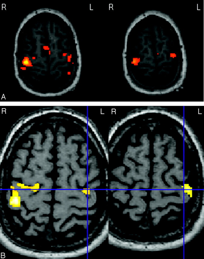
Axial functional MR imaging sequences showing the bilateral precentral cortical activation after motor tasks of the left hand in patients 14 (A) and 20 (B), amputated from the right upper limb (blue cross, enabling correlation between both images on B). This activation is obtained for analysis threshold corresponding to P values much greater than .0001. Minor differences are observed in surface and distribution of the activation between both sides.