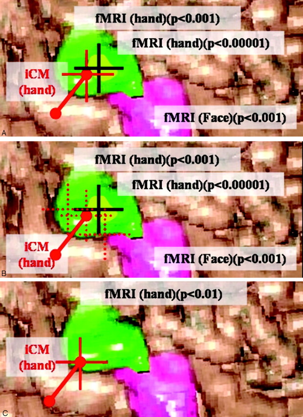
Fig 3.

Correlation, in the navigation system, between the iCM-defined targets (center of a 1-cm area between 2 poles of the grid but represented by a red cross) and the contours of the fMRI-defined activation areas (green and pink surfaces for hand and face, respectively, including focus of highest significance [centroid of the blob, black cross] designated as “fMRI target”) at the initial (or more restrictive) analysis threshold corresponding to P < .001 (or P < .0001). These pictures and the surface of cortical activation are only illustrative and do not represent actual data.
A, When targets are unambiguous (focal/reproducible/significant/with no artifact), we estimate that they correspond spatially only if the contours of the fMRI-activated area include the target of highest iCM wave.
B, When repeated iCM recordings provide ambiguous (diffused, not reproducible, altered by artifacts) results (red pointed square crosses), we designate as the iCM target the one defined by the recording presenting the highest amplitude (red cross). If this target is projected within the contours of the fMRI-activated area, we estimate that targets from both techniques corresponded spatially. When no iCM target is available, no comparison is possible.
C, When spatial concordance between both targets was obtained with lower thresholds than that corresponding to P < .001 (ie, when P < .01), we estimate that the concordance is not significant.
