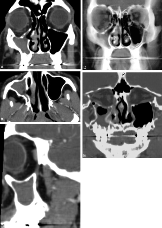
Fig 1.
(A) Coronal, (B) transverse, and (C) sagittal CT images of the sinuses show inward retraction of all walls of the right maxillary sinus with enlargement of the orbit and the middle meatus. The uncinate process is not clearly visualized because it is markedly thinned and retracted to the inferomedial orbital wall (confirmed with nasal endoscopy). (D) Thick-slab volume reconstruction in the coronal plane and (E) curved reconstruction along the optic nerves better demonstrates the maxillary sinus volume loss and enlargement of the orbit and middle meatus.

