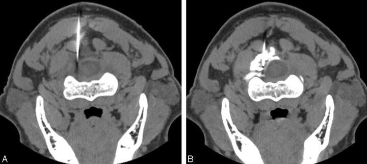
Fig 4.
Two axial CT images obtained with the patient in the prone position show needle placement in the left lateral epidural compartment at the upper C2 level (A) followed by administration of the blood patch; contrast material injected to confirm the epidural location is identified with mild flattening of the lateral thecal sac margin (B).

