Fig 1.
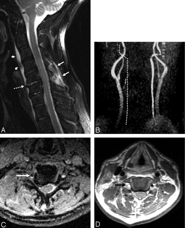
A 58-year-old man who sustained a unilateral interfacetal dislocation at C5–C6 without SCI.
A, Sagittal T2-weighted fast spin-echo (FSE) image (TR/TE/ETL: 2000/80/8) shows an injured disk at C5–C6 with increased signal intensity in the disk and probably avulsion of the anterior longitudinal ligament (dashed arrow). Prevertebral edema (arrowheads) and edema in the posterior paraspinal musculature (white arrows) are present.
B, Nonvisualization of the right vertebral artery. MIP image (anterior view) from a 2D time-of-flight acquisition (TR/TE/flip: 40/8.7/30) shows absence of signal intensity in the expected course of the right vertebral artery (dotted line).
C, Thrombus in the right foramen transversarium. Axial image from a 3D GRE acquisition (TR/TE/flip: 37/min/15) shows an oval area of low signal intensity in the right foramen transversarium corresponding to thrombus in the right vertebral artery. Note the normal flow-related enhancement in the left foramen transversarium.
D, Thombus in the right vertebral artery. Axial FSE image (TR/TE/ETL: 3000/28/4) obtained at a similar level to image in panel C shows a high-signal-intensity thrombus (arrow) in the right foramen transversarium indicative of a thrombosed vertebral artery. Note the normal flow void of the left vertebral artery in the left foramen transversarium.

