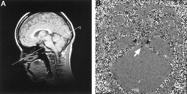
Fig 1.
Gradient recalled acquisition in the steady state and contrast-enhanced 2D cine phase MR images.
A, For the determination of the transverse image plane, a sagittal gradient recalled acquisition in the steady state MR image (20/5; flip angle, 60 degrees) was created. The imaging plane was determined at the midpontine level to be perpendicular to the basilar artery.
B, Contrast-enhanced 2D cine phase MR image (40/11.8; flip angle, 30 degrees), acquired at 32 phase. Note the basilar artery as an area of hypointensity (arrow).

