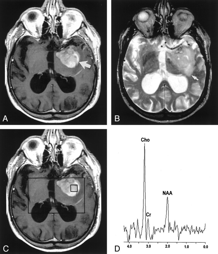
Fig 2.

Images in an 81-year-old man with a high-grade mixed glioma.
A, Contrast-enhanced axial T1-weighted image (600/14/1) demonstrates an ill-defined enhancing mass (arrow) in the left temporal region.
B, Axial T2-weighted image (3400/119/1) shows increased signal intensity (arrow) around the lesion.
C, Localizing image (600/14/1) for proton MR spectroscopy displays a voxel in the central portion of the lesion.
D, Proton MR spectrum obtained by using PRESS (1500/144) demonstrates an elevated Cho value and a decreased NAA value.
