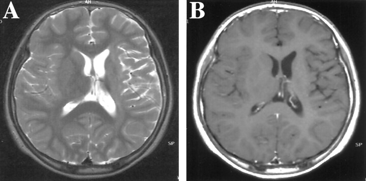
Fig 1.
MR imaging findings in the case of a 12-year-old, previously healthy and developmentally normal, left-handed Japanese male patient who developed slow progressive right-sided hemiparesis, which had originated in the right hand and gradually spread to the right leg during 1.5 years.
A, Axial view T2-weighted MR image (4000/96.0 [TR/TE]) shows a high intensity area in the left internal capsule, with volume loss of the left basal ganglia and thalamus. The lateral ventricle, sulci, and sylvian fissure are dilated on the left side.
B, Contrast-enhanced T1-weighted MR image (600/14) shows no enhancement in the lesion.

