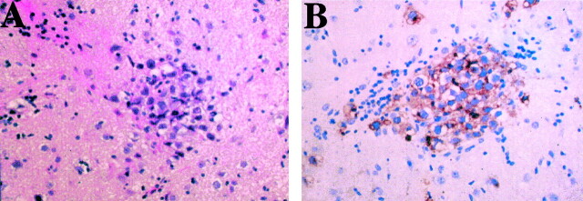
Fig 3.
Histopathologic examination of a specimen obtained from the left putamen by stereotactic biopsy.
A, Hematoxylin and eosin staining shows two different types of cells: large atypical cells (tumor cells) and infiltrating lymphocytes (original magnification, ×200).
B, Placental alkaline phosphatase staining shows positive tumor cells (original magnification, × 200)

