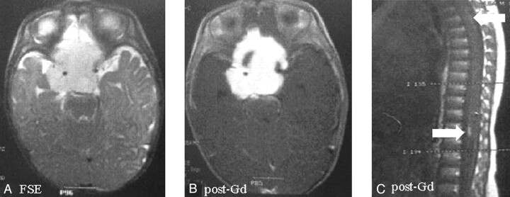
Fig 1.
Suprasellar pilomyxoid astrocytoma in a 5-month-old boy (patient 2). The mass, surrounding the circle of Willis, shows hyperintense signal on fast spin-echo images (A) and homogeneous enhancement after contrast material administration except for right anterior small cystic component (B). Peripheral pial enhancement of spinal cord on postcontrast sagittal T1 spin-echo images was detected (C).

