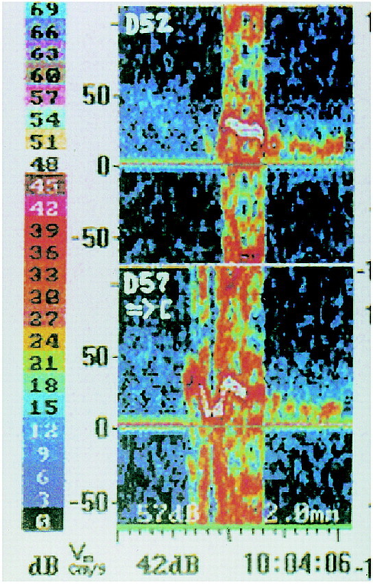
fig 1.

The TCD device provides two sample volumes for insonation of the MCA. An embolus streaming down the MCA will be recorded first in the proximal sample volume (depth of insonation, 57 mm [D57]) and with a time delay in the distal sample volume (depth of insonation, 52 mm [D52]). Next to the color-coded decibel scale, the Doppler velocity spectrum after fast Fourier transformation (FFT) of each sample volume is shown. The blood flow direction in both sample volumes is indicated in the proximal sample volume beneath D57 by an arrow directed toward the probe symbol ([), which means that the velocity spectrum of the blood flow in the MCA usually directed toward the probe is imaged above the zero line; the background velocity spectrum can be seen more clearly in the distal sample volume as a band with a signal intensity of 9 to 15 dB (colored slightly blue to green). The signal of the embolus after FFT is overloaded, as indicated by its bidirectional appearance
