fig 2.

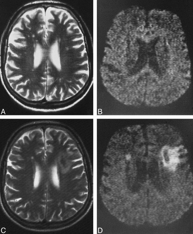
A–D, Pre- and postoperative T2-weighted images (A and C) and DWIs (B and D) of a patient in whom a territorial infarct was present on the postoperative images. The small white matter lesions on the preoperative T2-weighted (4000/99/3) image (A) are not present on the preoperative DWI (B) (episequence; 123/1 [TE/excitations]). The corresponding postoperative T2-weighted image (C) and DWI (D) show a medium-sized territorial infarct on the left side, and a small new lesion in the head of the caudate nucleus on the right side. This patient underwent surgery for a tight left-sided stenosis associated with occlusion of the contralateral internal carotid artery
