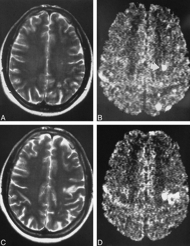
fig 4.

A–D, Pre- and postoperative images of a patient who underwent carotid endarterectomy 14 days after the clinical event. The preoperative T2-weighted (A) and DWI (B) studies show four small cortical lesions and one subcortical lesion (arrow, B). The cortical lesions cannot be detected on the corresponding postoperative DWI (D). The subcortical lesion is still present, but there is a new, smaller territorial infarction nearby as shown on the T2-weighted (C) and DWI (D) studies. These findings may suggest that cortical lesions disappear sooner than subcortical lesions, probably because of the better collateral blood flow in cortical regions as compared with subcortical regions
