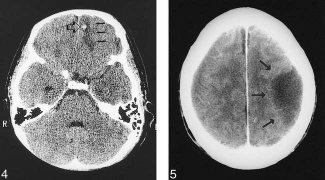
fig 4.

15-year-old girl with left hemotympanum. An emergency temporal bone CT was performed to evaluate for temporal bone fracture. The initial interpretation by the on-call resident identified a left temporal bone fracture. The next morning, the staff neuroradiologist also noted a hemorrhagic contusion of the left frontal lobe (open arrow), with surrounding edema (closed arrows). This error in interpretation was given a grade 2 (would not ordinarily be expected to be identified) by the panel. The patient was called back for reevaluation and a follow-up head CT. The 11-hour delay in diagnosis did not change her outcome.fig 5. 70-year-old man with metastatic prostate cancer, presented with right-sided weakness and mental status changes. An emergency head CT was performed to evaluate for metastasis or infarct. The initial interpretation by the on-call resident was intra-axial edema (arrows) from a metastasis or infarct. The next morning, the staff neuroradiologist interpreted this lesion to represent an extra-axial fluid collection. This error in interpretation was given a grade 2 (would not ordinarily be expected to be identified, difficult to distinguish) by the panel. The initial error in interpretation resulted in a 13-hour delay in treatment, as the patient was taken to the operating room for drainage of the subdural hematoma the next day; however, his outcome was not changed
