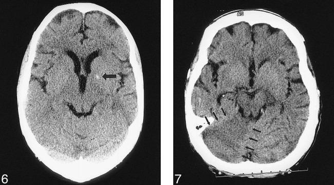
fig 6.

66-year-old man with right hemiparesis and aphasia. An emergency noncontrast head CT was performed to determine whether the patient was a potential candidate for thrombolysis. The initial interpretation by the on-call resident identified a high-attenuation focus in the left basal ganglia (arrow), most likely representing hemorrhage. The next morning, the staff neuroradiologist interpreted this lesion as calcification, not hemorrhage. This error in interpretation was given a grade 3 (could usually be identified) by the panel. The initial misinterpretation resulted in thrombolytic therapy being withheld, denying the patient the potential benefit of that treatment and thereby representing a potentially serious change in his outcome.fig 7. 61-year-old woman with end-stage renal disease presented with a change in mental status. An emergency noncontrast head CT was performed to evaluate for septic emboli or hemorrhage. The initial interpretation by the on-call resident was negative. The next morning, the staff neuroradiologist noted a right cerebellar infarct (arrows). This error in interpretation was given a grade 3 (could usually be identified) by the panel. The initial misinterpretation resulted in a change in management, as the patient received dialysis to exclude uremia as the possible cause of her symptoms, and a delay in the treatment of her stroke, resulting in a potentially serious change in the patient's outcome
