fig 2.

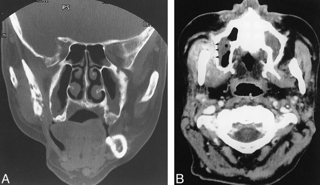
Images of a 49-year-old man treated for right-sided soft palate squamous cell carcinoma.
A, Coronal CT scan (bone window) shows cortical disruption and fragmentation of the right ramus.
B, Contrast-enhanced CT scan shows prominent thickening and enhancement of the adjacent masticator muscles. The buccal space is also involved (arrowheads).
