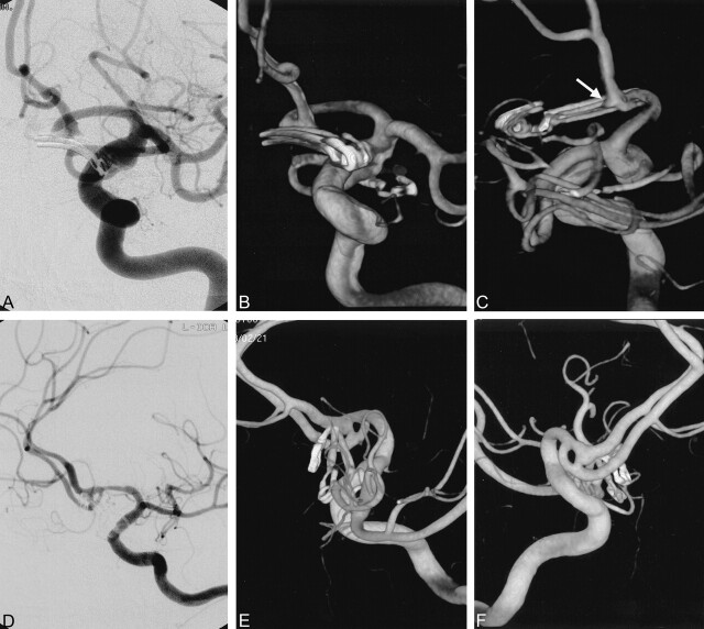
Fig 2.
Small, dog-eared remnant aneurysm.
A—C, Case 24. Postoperative DSA (A) performed 8 days after surgery for anterior and posterior communicating artery aneurysms shows no residual. Anterior 3D angiogram (B) shows the vessels and clips in the two aneurysms well. Posterior 3D angiogram (C) reveals a tiny, dog-ear remnant in the anterior communicating artery region (arrow).
D–F, Case 5. Postoperative DSA (D) of a clipped anterior communicating artery aneurysm shows no definite residual. Anterior oblique and posterior oblique 3D aneurysms (E, F) reveal a residual aneurysm near the ends of the clips and its relationship with a fenestration.

