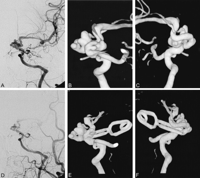
Fig 4.
Case 65. Dolichocarotid aneurysms.
A–C, Anterior and posterior preoperative DSA and 3D angiographic views show a complex aneurysms involving the distal ICA.
D, On postoperative DSA, it is hard to see which part of the aneurysms is clipped.
E and F, Anterior and posterior 3D angiograms reveal that the lateral part of the aneurysms is clipped, and part of ICA is included within the end of the clip.

