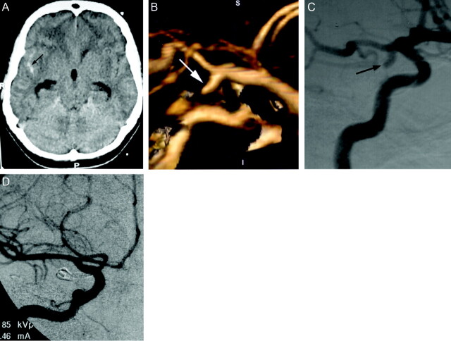
Fig 1.
Images obtained in a 45-year-old woman with severe headache.
A, Unenhanced CT scan of the head shows subarachnoid hemorrhage in the right sylvian fissure (arrow), with mild hydrocephalus. R, right; P, posterior.
B, 3D volume-rendered image (lateral) shows inferiorly and posteriorly directed saccular aneurysm at the origin of the right posterior communicating artery (arrow). S, superior; I, inferior; P, posterior; A, anterior.
C, Preoperative right internal carotid digital subtraction angiogram (right anterior oblique projection) shows inferiorly and laterally directed saccular aneurysm at the origin of the posterior communicating artery (arrow).
D, Intraoperative right internal carotid digital subtraction angiogram (anteroposterior projection) shows successful clip placement in the posterior communicating artery aneurysm.

