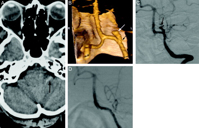
Fig 2.
Images obtained in a 49-year-old woman with severe headache.
A, Unenhanced CT scan of the head shows subarachnoid hemorrhage in the left cerebellopontine angle cistern (arrow).
B, 3D volume-rendered image (posteroanterior) shows superiorly, medially, and anteriorly directed saccular aneurysm (white arrow), which is incorporated into the origin of posterior inferior cerebellar artery (black arrow). S, superior; I, inferior; R, right; L, left.
C, Preoperative left vertebral artery digital subtraction angiogram (anteroposterior projection) shows saccular aneurysm projecting superiorly and medially at the origin of the posterior inferior cerebellar artery. Note the hypoplastic P1 segment of the posterior cerebral artery.
D, Intraoperative left vertebral artery digital subtraction angiogram (anteroposterior projection) shows successful clip placement in the aneurysm without occlusion of the posterior inferior cerebellar artery.

