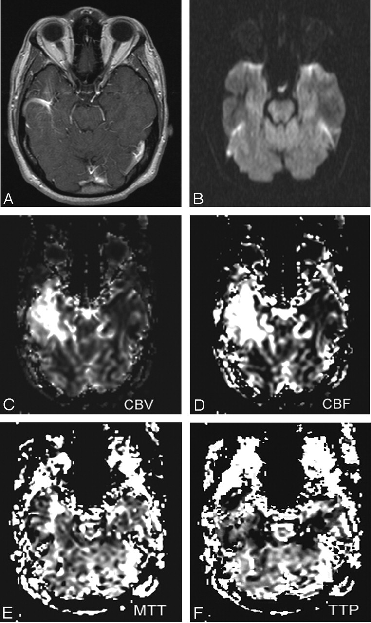
Fig 2.

Axial T1-weighted postcontrast image (TR/TE, 635/17) (A), axial DW image (TR/TE, 4300/122; b = 1000) (B), and axial gradient echo CBV (C), CBF (D), MTT (E), and TTP (F) maps of the brain in a patient with an uncomplicated DVA (case 3). The postcontrast image demonstrates a DVA in the right temporal lobe. DW image demonstrates flow void in the draining vein of the DVA, but no restriction of diffusion. Perfusion maps show marked elevations in CBF and CBV and milder elevations in MTT and TTP within the DVA and in the surrounding parenchyma.
