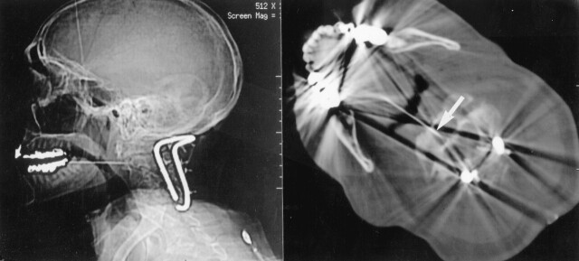
Fig 5.
Lateral scout and axial CT images through C2 show a transoropharyngeal approach to a lesion in the body of C2 in a patient without a history of cancer but who was first stabilized posteriorly. Arrow shows the needle tip in the C2 lytic lesion. Biopsy showed squamous cell carcinoma, possibly from the lung or upper aerodigestive tract. The primary site was never determined.

