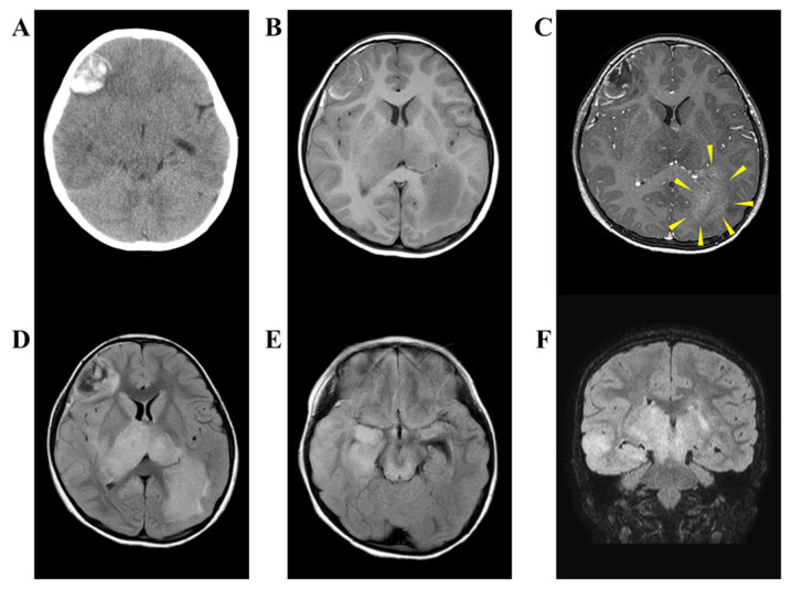
Figure 1.
Preoperative CT and MRI. (A) Plain CT shows acute phase hemorrhage of the right frontal lobe. (B) In T1-weighted image of MRI, the frontal lobe lesion shows a low intensity and its margin has a high intensity. Bilateral thalamus and left occipital lobe also show low intensity. (C) Edge of the lesion is enhanced by gadolinium. Reticulated enhanced effect is observed in left occipital lobe (arrow head). (D-F) In the fluid-attenuated inversion recovery MRI, the frontal lesion shows a patchy inside and its margin exhibit a high signal. The bilateral thalamus, right temporal, and left occipital exhibit high intensity and they are continuous. CT, computed tomography; MRI, magnetic resonance image.

