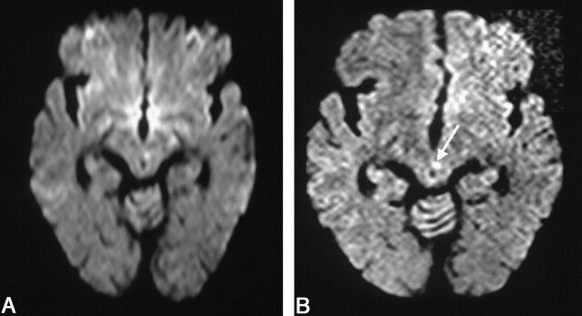
Fig 1.
Images in a 67-year-old man with oculomotor nerve palsy.
A, Conventional DW imaging fails to show any significant abnormality.
B, The thin-section DW imaging shows a tiny hyperintense lesion at the left paramedian midbrain (arrow). The TOAST diagnosis was changed from normal to small-vessel disease.

