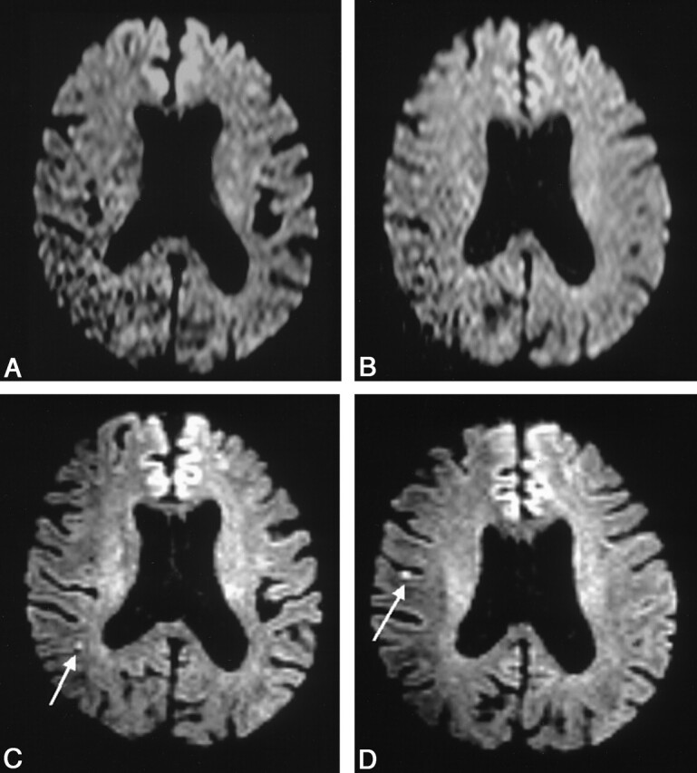
Fig 2.

Images obtained in a 67-year-old man with aphasia.
A and B, Conventional DW images do not depict any lesions.
C and D, Thin-section DW images depict lesions in the cerebral hemisphere in the middle cerebral artery territory (arrows). The TOAST diagnosis was changed from normal to large-artery atherosclerosis.
