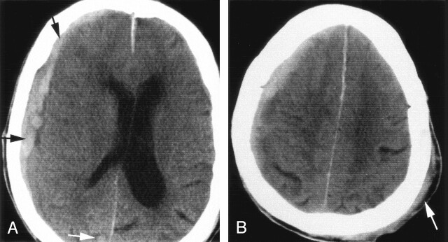
Fig 1.
Images obtained in a 93-year-old woman treated with coumadin for atrial fibrillation. She fell and hit her head at home and presented with nausea, vomiting, a positive left-sided Babinski reflex, and a GCS score of 13.
A, Axial nonenhanced CT scan reveals acute right frontoparietal subdural hematoma (black arrows) with a moderate degree of midline shift. Image also shows a small occipital hemorrhagic contusion on the right (white arrow).
B, More cephalic CT scan shows a soft tissue hematoma on the left (arrow) consistent with a contrecoup traumatic subdural hematoma. The patient’s anticoagulation status was reversed with vitamin K and fresh frozen plasma. The patient’s clinical condition deteriorated further. She was not a neurosurgical candidate and therefore given conservative care at the request of her family.

