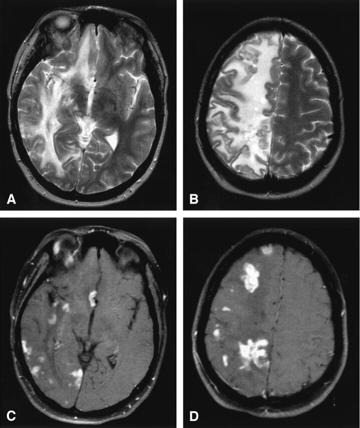
Fig 1.

Lymphocytic vasculitis, MR imaging.
A and B, Axial T2-weighted MR images reveal multifocal regions of T2 hyperintensity with surrounding diffuse vasogenic edema limited to the right cerebral hemisphere resulting in mild right to left midline shift.
C and D, Axial T1-weighted postgadolinium MR images with fat saturation depict multifocal enhancing tumefactive lesions, primarily at the subcortical gray-white matter junction and basal ganglia, throughout the right cerebral hemisphere with sparing of the left cerebral hemisphere.
