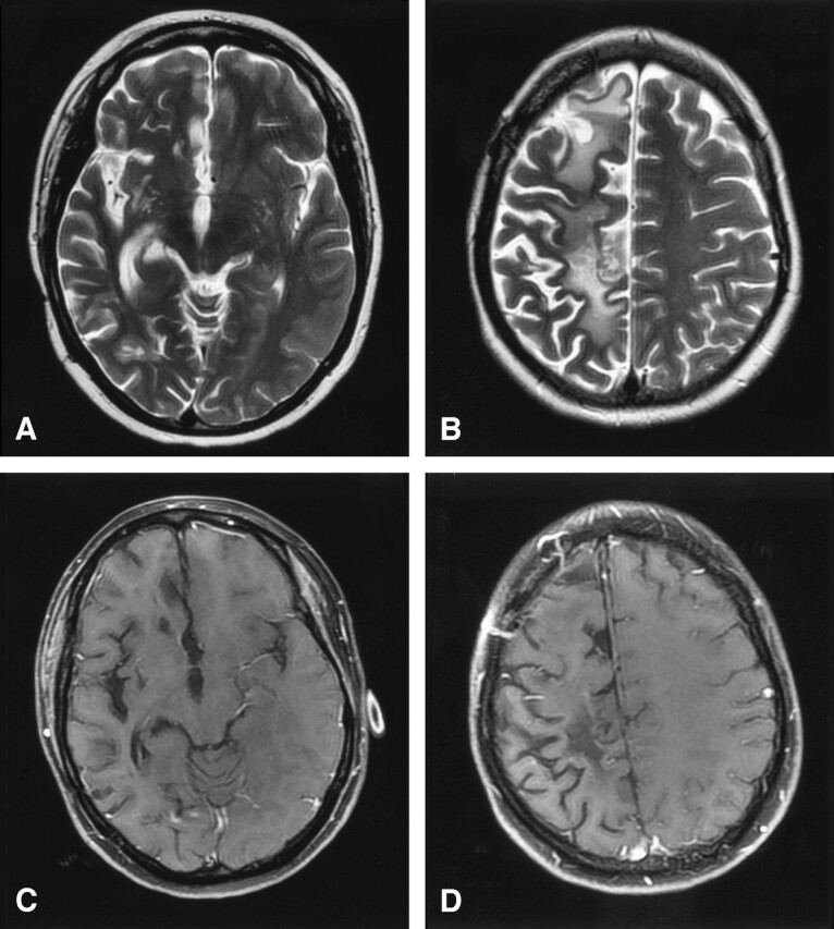
Fig 4.

Lymphatic vasculitis, MR imaging post-treatment.
A and B, Axial T2-weighted images 5 months after therapy show resolution of edema and mass effect with residual foci of gliosis and extensive atrophy of the right cerebral hemisphere.
C and D, Axial T1-weighted postgadolinium images with fat saturation demonstrate interval resolution of the previously seen enhancing masses. Craniotomy defect is evident in the right frontal region from the previous biopsy.
