Fig 5.
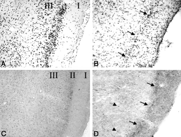
Cresyl violet (A and B) and silver impregnation (C and D) stains of the piriform cortex (magnification ×10).
A, Sections in a control rat after imaging shows no loss of neurons. Note the density of darkly stained neuronal nuclei in layers II and III.
B, Seven days after the induction of seizures, marked neuronal loss and vacuolization of layers II and III are seen. Note the decreased neuronal density of layer II (arrows).
C, Section in a control animal confirms the lack of neuronal loss.
D, Few argyrophilically stained neurons (arrowheads) are visible deep in the piriform cortex 7 days after seizure induction, because most degeneration took place earlier. Note the loss of neurons in layer II (arrows) and vacuolization of layer III.

