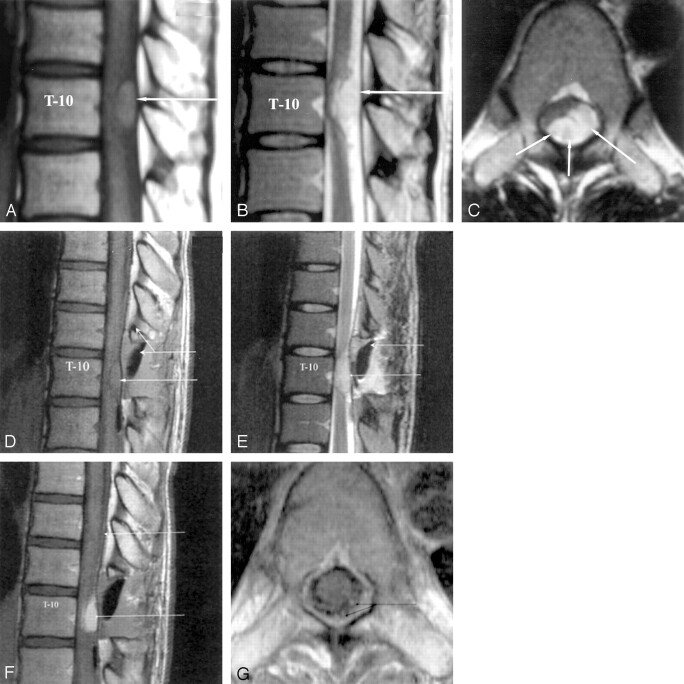
Fig 1.
Sagittal and axial MR images.
A and D, T1-weighted sagittal images, showing an intradural extramedullary lesion at T10 that is isointense relative to cord (single arrow). Preoperative appearance (A) and postoperative(D) changes are shown (double arrows).
B and E, Lesion is hyperintense on T2-weighted sagittal images (arrow). Postoperative changes are shown (large arrow, E).
C, Postcontrast T1-weighted axial image, showing intense enhancement of lesion (arrows).
F, Postcontrast T1-weighted sagittal image, showing intense tumor enhancement (lower arrow) and cranially draining dorsal enhancing perimedullary veins (upper arrow).
G, Postcontrast T1-weighted axial image showing enlarged enhancing draining veins on the dorsal surface of the cord above the lesion (arrows).

