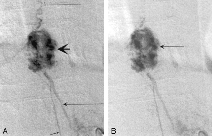
Fig 2.
Spinal arteriogram.
A, Early arterial phase image, showing a hypervascular nodule (large arrowhead) that demonstrates predominantly peripheral lobular inhomogeneous enhancement. Feeder from the left T11 intercostal artery (large single arrow), radiculomedullary feeder to the cord (single small arrow), and draining veins (double arrows) are seen.
B, Late arterial phase image, showing that the tumor nodule enhances more homogeneously (arrow).

