
Fig 1.
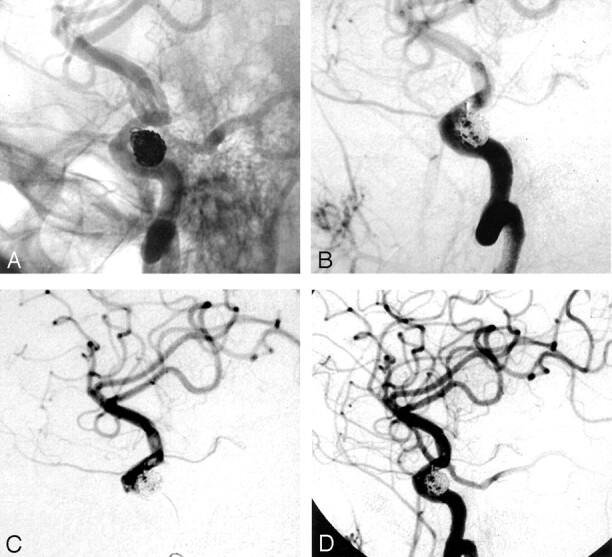
Illustrative case 1.
A, Right internal carotid artery angiogram, oblique projection, unsubtracted, shows a coil loop protruding into the parent vessel.
B, Right internal carotid artery angiogram, oblique projection, subtracted view, shows a filling defect about the coil loop, indicating thrombus formation. Note the decreased antegrade flow into the supraclinoid internal carotid artery.
C, Right internal carotid artery angiogram obtained via a microcatheter injection. The image clearly shows the filling defects about the protruded coil loop and more distally.
D, After a local, low-dose (2-mg), intra-arterial infusion of abciximab through the microcatheter, thrombus dissolution is documented without evidence of distal thromboembolism.