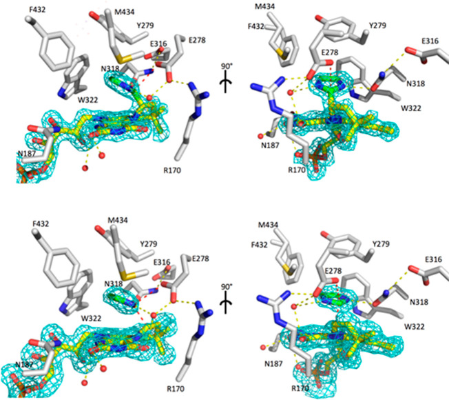
Figure 13.
PA0254UbiX active site structure. Active site residues shown in atom-colored sticks (gray carbons) with the prFMNiminium cofactor shown with yellow carbons. Bound imidazole derived from the crystallization buffer is shown with green carbons. Omit FoFc map corresponding to cofactor and imidazole is contoured at 3 sigma and shown as a cyan mesh. In two monomers a covalent bond is formed between imidazole C2 and the prFMN C1′ (top view), while the active site of the other monomers lacks electron density in between the imidazole and the prFMN, indicating a noncovalent complex. Hydrogen bonds are shown in yellow dotted lines, while the key Glu278 imidazole C2 and imidazole C2/prFMN C1′ interactions are shown in red.

