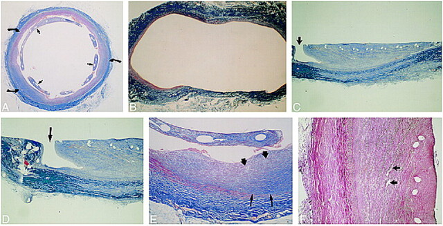
fig 4.
Histopathologic findings.
A, Mason's trichrome stain on stented aneurysm shows the aneurysmal lumen is nearly completely filled in with organized fibrous connective tissue (curved arrows). The stent wires are surrounded by neointima (straight arrows).
B, Mason's trichrome stain on control aneurysm in cross section shows markedly distended thin wall of the aneurysm.
C and D, Trichrome stain on stented aneurysm in longitudinal section shows a small, patent endothelial-lined channel at one margin, connecting the carotid lumen with the aneurysmal lumen (arrow). These small channels were seen in all aneurysmal specimens. The remainder of the aneurysmal lumens were filled in by fibrous connective tissue (organized thrombus). Within the neointima surrounding the stent wires there was mild inflammatory cell infiltration (macrophages). Many macrophages contained blood pigment (resolving thrombus).
E, Magnified cross-sectional view of aneurysmal lumen with Mason's trichrome stain. Fibrin and collagen stain blue. Dark blue represents more mature fibrin and collagen. Mature fibrous elements and collagen (dark blue) were noted near the periphery of the aneurysmal wall (thin arrows); less mature fibrous elements (light blue) lie near the central lumen of the aneurysm (thick arrows), suggesting that thrombus organization begins along the outer wall and progresses in a centripetal fashion toward the central lumen of the aneurysm.
F, Verhoeff-van Gieson elastic stain shows heavily vascularized organized thrombus (arrows).

