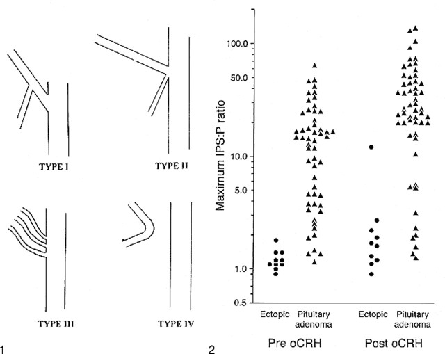
fig 1.
Modified classification of IPS anatomic variants. Type I consists of an IPS anastomosing with the internal jugular vein. The anterior condylar vein is absent or joins the IPS at a defined origin. The short segment of vein from the point of this anastomosis to the internal jugular vein is termed the inferior condylar confluence. Type II consists of a common origin of the IPS and anterior condylar vein with the internal jugular vein. Types III and IV remain as originally described by Shiu et al (5) (ie, an IPS consisting of several small channels communicating with the internal jugular vein and an IPS that communicates with the anterior condylar vein and not the internal jugular vein, respectively).fig 2. Maximum IPS-to-peripheral-blood ACTH ratios before and after oCRH administration in 65 procedures in which confirmation of the ACTH source was obtained

