fig 2.
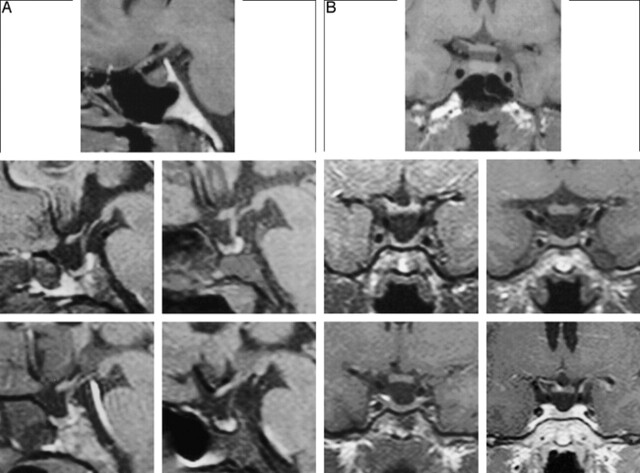
Fat-suppressed T1-weighted MR images of four adult male dwarfs show the pituitary area affected by a mutation in the GHRH receptor. An MR study of a normal 25-year-old man is shown at the top for comparison. The four lower panels show, in sequence (upper left to lower right), MR images of four patients (ages, 22, 27, 27, and 29 years, respectively). Note the hypoplastic anterior pituitary and the normal posterior lobe. As a result of adenohypophyseal hypoplasia, the neurohypophyseal “bright spots” appear very prominent. The MR image of the patient shown in figure 1 is on the upper left.
A, Sagittal views. All images were obtained without the administration of contrast material.
B, Coronal views. The coronal image on the lower right was obtained after the administration of contrast material; all others were obtained without the administration of contrast material.

