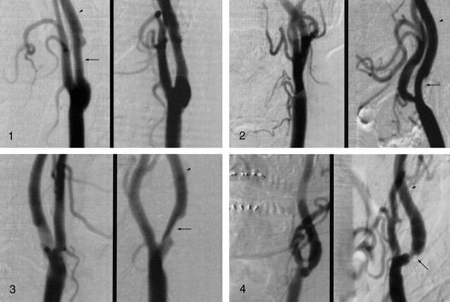
fig 1.
Results of Doppler sonography of the carotid arteries were interpreted as mild stenosis with a peak systolic velocity of 104 cm/s. Percent stenosis was measured as 25% according to the original interpretation and as 23% by the blinded readers. Arrow indicates point of maximum stenosis, and arrowhead indicates normal distal internal carotid artery.fig 2. Results of sonography of the carotid arteries were interpreted as moderate stenosis with a peak systolic velocity of 132 cm/s. Percent stenosis was measured as 33% according to the original interpretation and as 28% by the blinded readers. Arrow indicates point of maximum stenosis, and arrowhead indicates normal distal internal carotid artery.fig 3. Results of Doppler sonography of the carotid arteries were interpreted as severe stenosis with a peak systolic velocity of 170 cm/s. Percent stenosis was measured as 70% according to the original interpretation and as 52% by the blinded readers. Arrow indicates point of maximum stenosis, and arrowhead indicates normal distal internal carotid artery.fig 4. Results of Doppler sonography of the carotid arteries were interpreted as critical stenosis with a peak systolic velocity of 450 cm/s. Percent stenosis was measured as 83% according to the original interpretation and as 61% by the blinded readers. Arrow indicates point of maximum stenosis, and arrowhead indicates normal distal internal carotid artery

