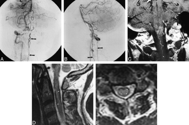
Fig 4. A and B, Late arterial-phase anteroposterior (A) and lateral (B) right vertebral artery injection shows filling of dural arteriovenous fistula (open arrow, B) originating from intradural vertebral artery. Drainage along cervical spinal cord is visible in both views (closed arrows).
C, Coronal contrast-enhanced MR image shows intraaxial cervicomedullary enhancement and enlarged pial cervical vein (arrowheads).
D and E, Sagittal (3733/100eff) (D) and axial (4000/100eff) (E) T2-weighted images of cervical cord show subpial hypointensity (arrowheads).

