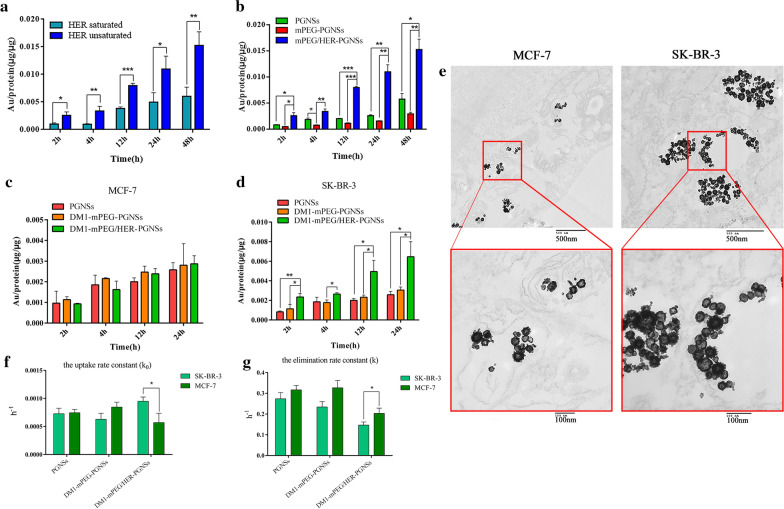
Fig. 5.
a Quantitative comparation of mPEG/HER-PGNSs internalized into SK-BR-3 cells with or without Her-2 antibody blocking; b Quantitative comparation of PGNSs, mPEG-PGNSs and mPEG/HER-PGNSs internalized into SK-BR-3 cells at 2, 4, 12, 24 h and 48 h using ICP-MS; c Quantitative comparation of PGNSs, DM1-mPEG-PGNSs and DM1-mPEG/HER-PGNSs internalized into MCF7 cells and d SK-BR-3 cells at 2, 4, 12, and 24 h using ICP-MS; and e TEM thin sections of cellular uptake of DM1-mPEG/HER-PGNSs in MCF7 cells and SK-BR-3 cells after 12 h (Scale bar = 500 nm, 100 nm); f k0 and e k values for cellular uptake and elimination in SK-BR-3 cells and MCF-7 cells. (*p < 0.05,**p < 0.01,***p < 0.001)

