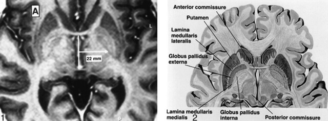
fig 1.
Axial FSE-IR image [3000/40/4 (TR/TE/excitations), with TI = 200 ms, echo train length = 5, 2 mm slice thickness] through the basal ganglia. Conventional localization of the GPi is a point 22 mm lateral to the midportion of the AC-PC line. This approach does not take into account the normal variations in laterality of the GPi.
fig 2. Anatomy of the GP. Axial diagrammatic depiction of the GPi from the GPe. The “lamina medullaris medialis” is synonymous with the GPi-GPe lamina (Borrowed with permission from Cohn and colleagues. Pre- and postoperative MR evaluation of stereotactic pallidotomy. AJNR 1998;19:1075–1080.).

