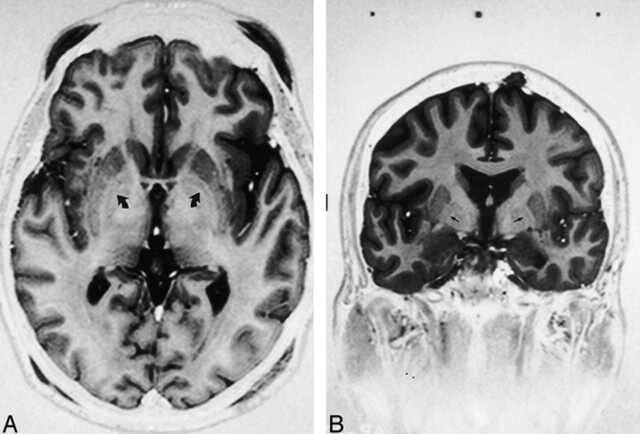
fig 3.
Axial (A) and coronal (B) fast spin-echo inversion-recovery sequences [3000/40/2–4, TI = 200 ms, echo train length = 5, 2-mm slice thickness] show GPi-GPe lamina (small arrows), separating the GPi from the GPe. On the axial image, the posterior commissure is not seen. The frame placement technique described only approximates the AC-PC line

